
CBCT
Cone Beam CT scanner
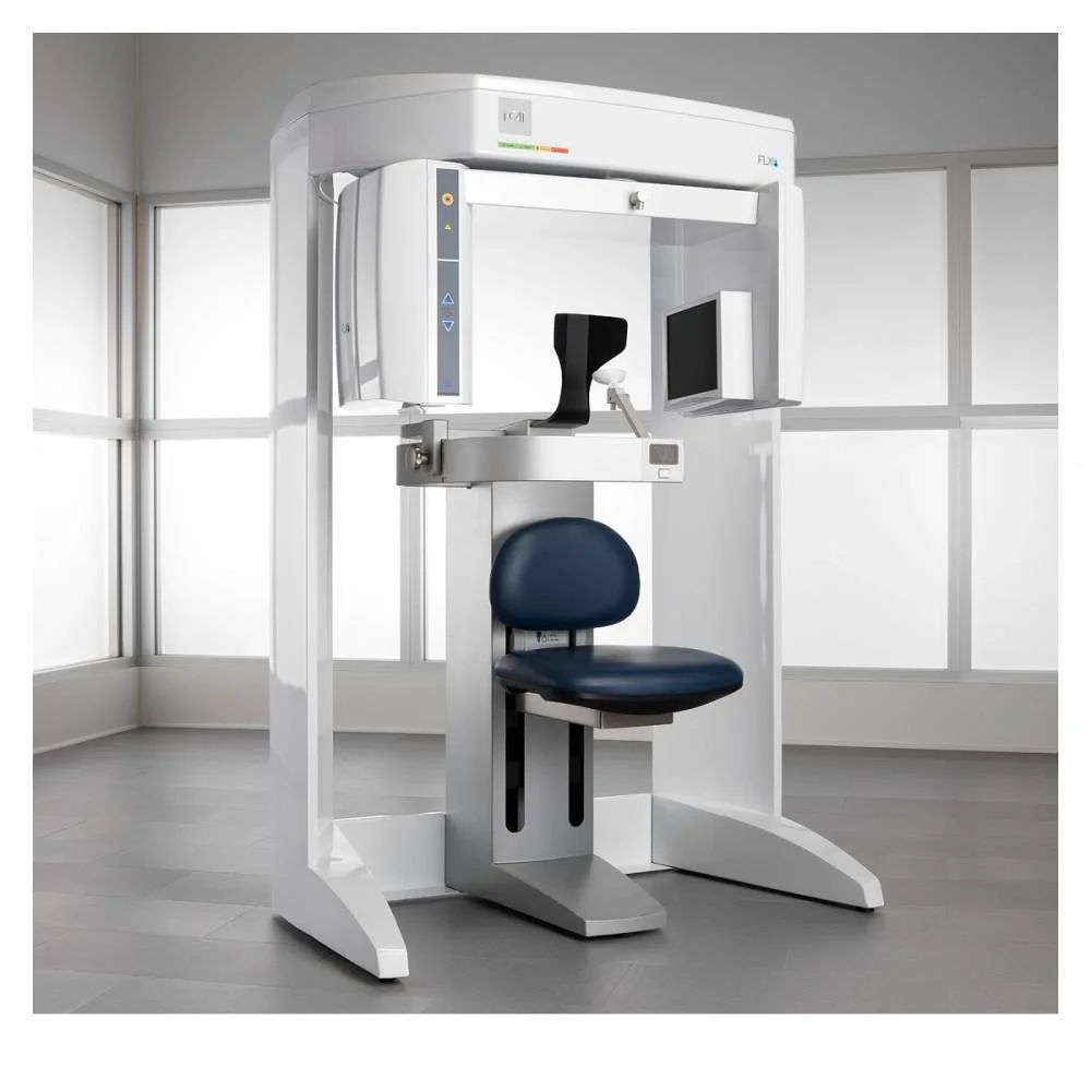
3D cone beam computed tomography (CBCT) is an imaging technology that allows us to better evaluate your face and jaws. It is different from a panoramic x-ray, which also rotates around your head, but is 2-dimensional. The 3-dimensional image produced by a CBCT scanner reveals underlying skeletal anatomy, anatomic location of nerves, exact position of impacted teeth, and so much more. It has made the field of oral and maxillofacial surgery a whole lot safer for patients, because we are able to have a better map of your anatomy before any surgery.
It has helped to revolutionize the field of 3-dimensional treatment planning for reconstructive jaw surgery and cleft surgery. Using a CBCT scan, we can virtually plan surgeries in advance in 3 dimensions, and have more accurate predictions of the result.
CBCT scan gives us a similar amount of information as a medical CT scan, but uses only a fraction of the radiation.
Virtual Surgical Planning in Orthognathic Surgery
VSP
CBCT technology allows us to see the bony anatomy in three dimensions. We can then virtually plan complex orthognathic (jaw) surgery in three dimensions. This is groundbreaking, because previously, we were limited to planning a 3 dimensional procedure in only two dimensions. Now, we have the ability to make virtual surgical movements in the X, Y, and Z axes, and see how each one affects the final result.
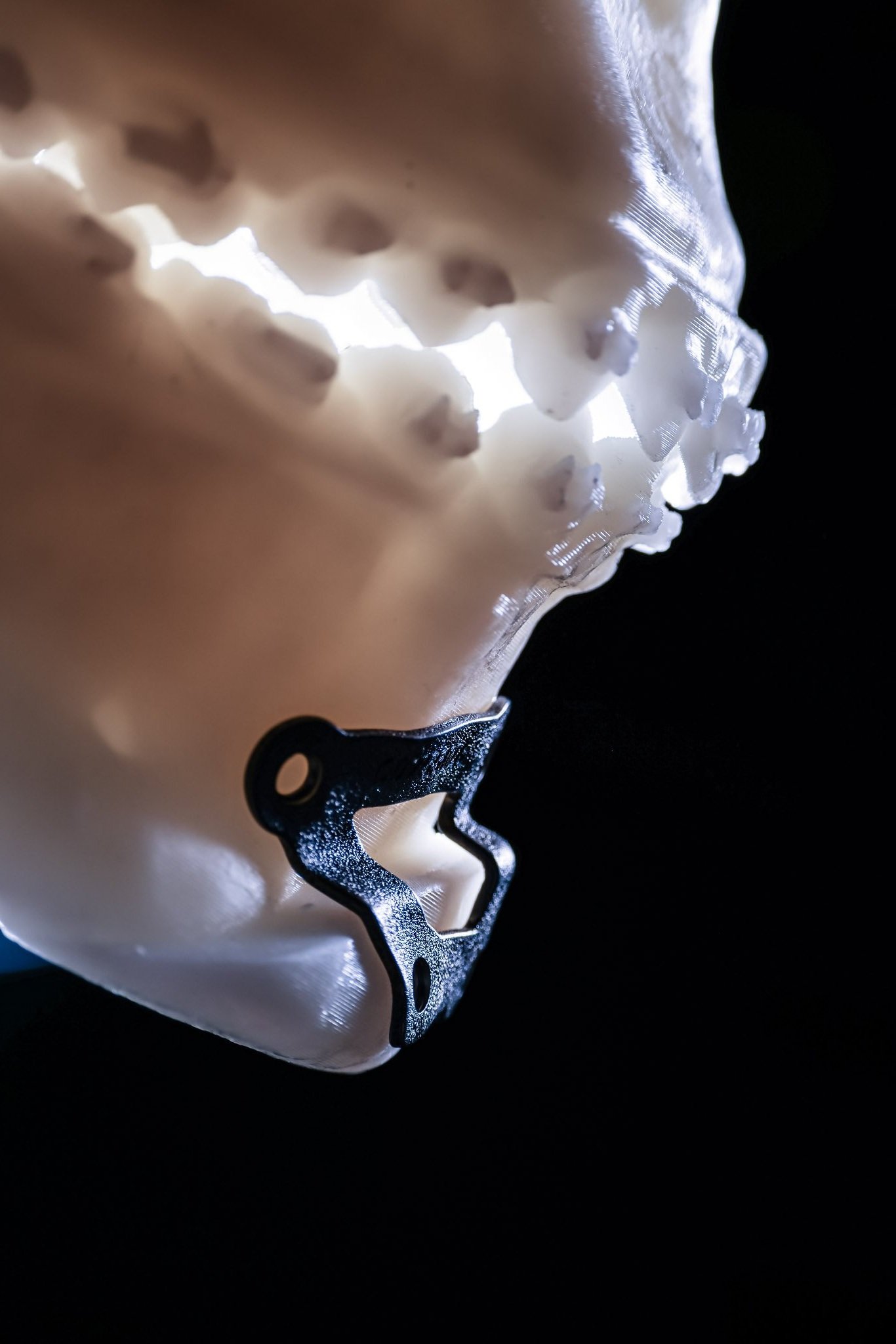
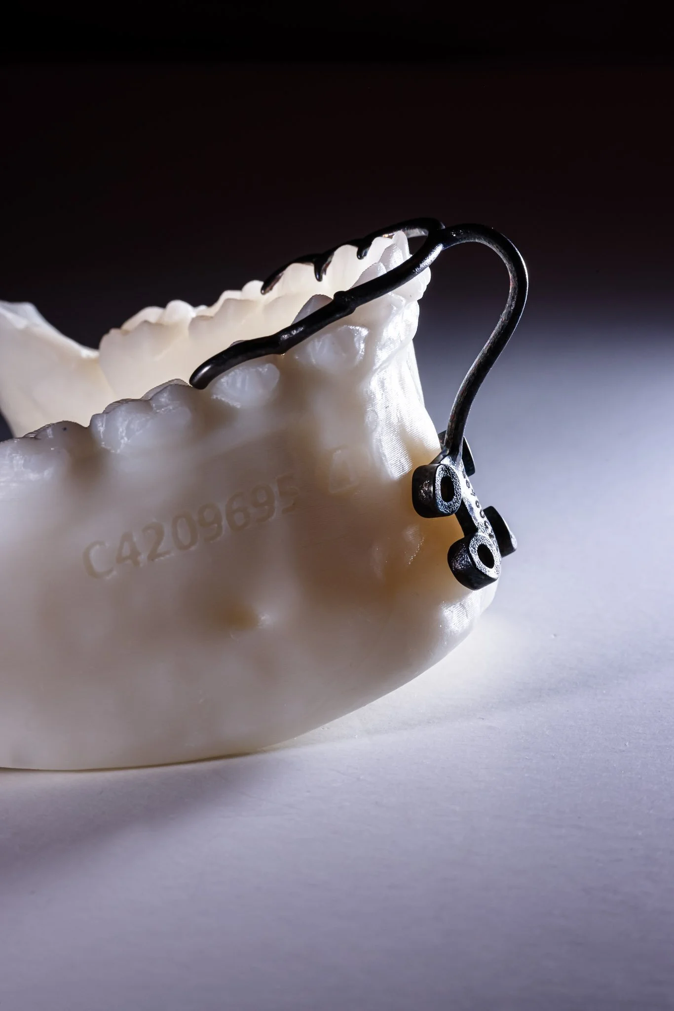
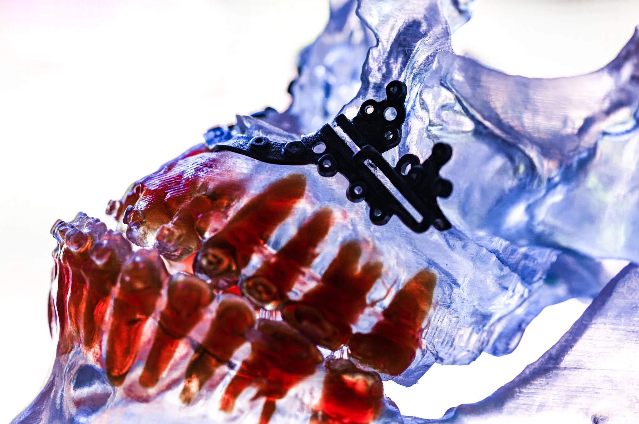
3D Printing in Oral and Maxillofacial Surgery
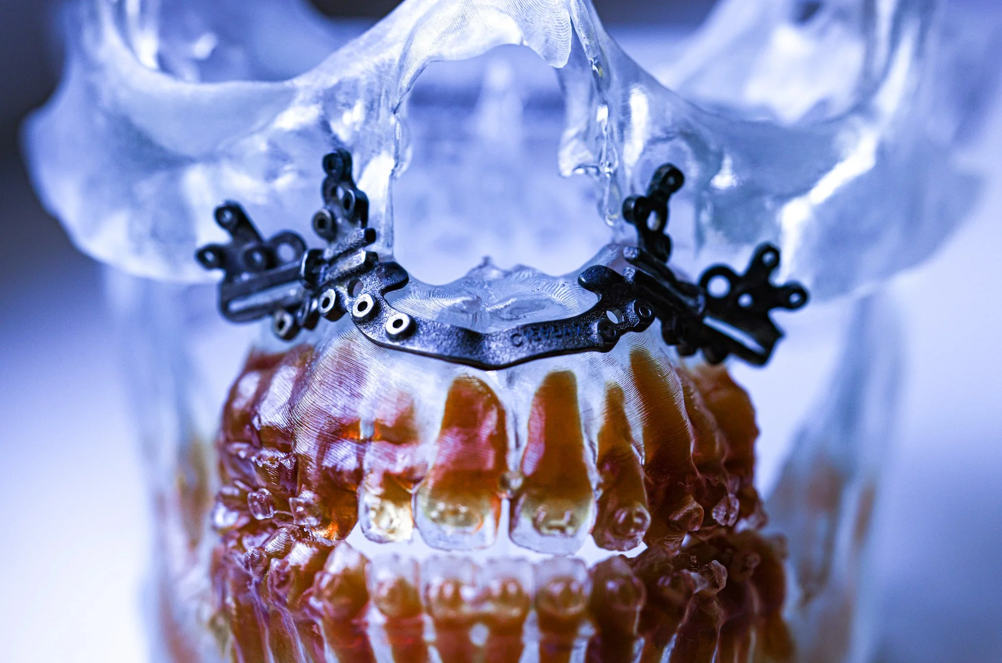
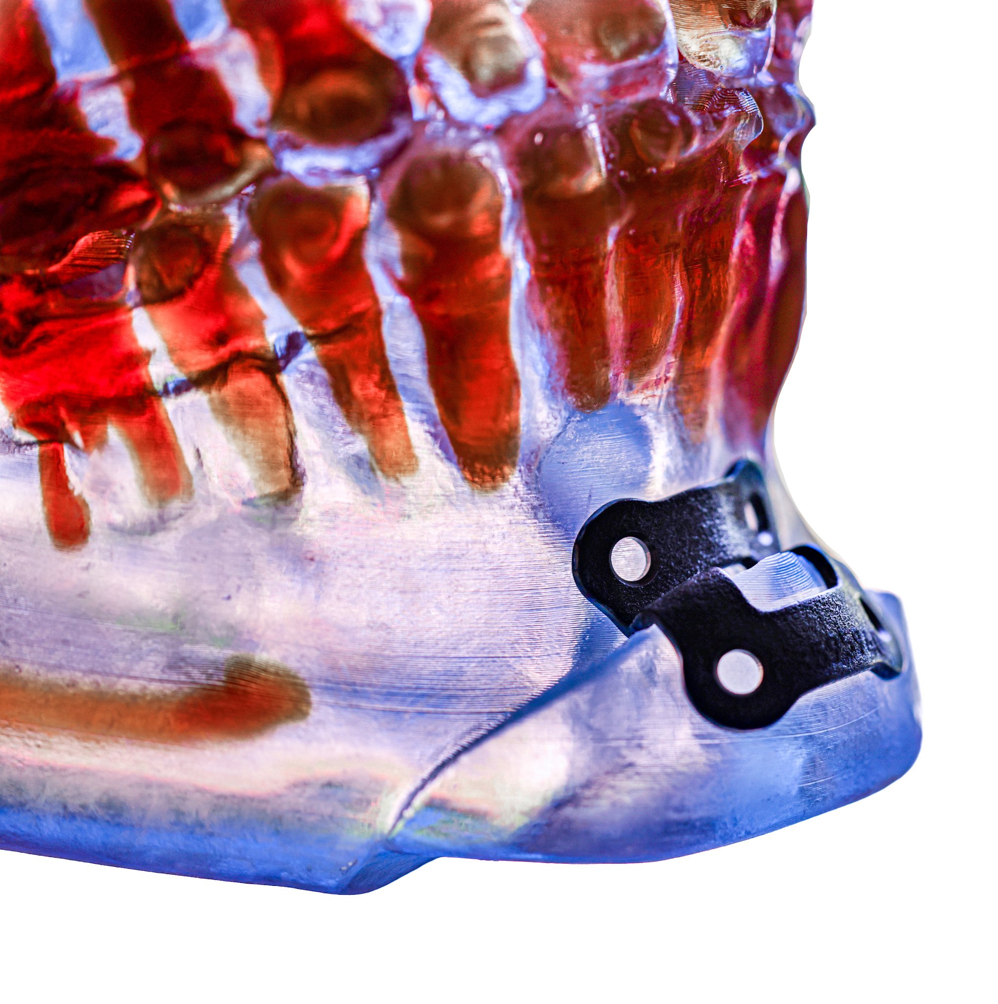
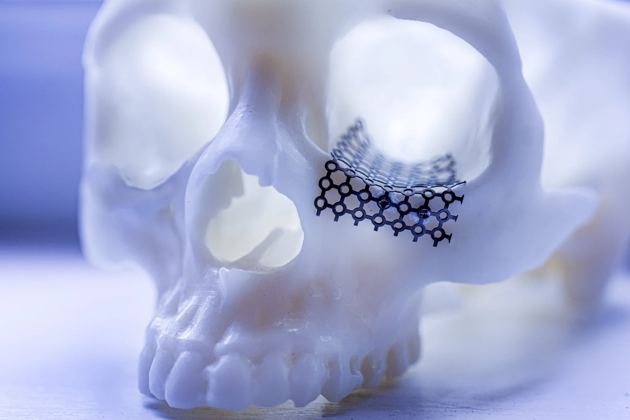
Taking the three dimensional virtual surgical planning a step further, we are now able to design custom titanium guides and custom titanium hardware in order to achieve a surgical result that is more accurate than was ever possible before. We are talking accuracy down to the fraction of a millimeter. The custom 3D printed titanium guides hug every curve of the bone anatomy and fit like a glove. This means more accurate surgical results, shorter surgical time, and stronger plates holding your bones. It’s win-win-win.

We have a three-dimensional intraoral scanner that allows us to scan the intricate surface anatomy of the teeth and surrounding structures. It also allows us to 3D print any appliances or guides that register to the teeth. It has revolutionized treatment with more accurate placement of dental implants, as well as more accurate guides for jaw surgery. We also use this technology to 3D print custom night guards for teeth grinding and TMJ pain.
Intraoral Scanner
3D printed custom hardware for orthognathic surgery
3D printed custom nightguard

Custom hardware for facial fractures
Platelet Rich Fibrin

Platelet Rich Fibrin, or PRF, allows the body to heal at an accelerated rate. When our bodies heal, platelets are the first line of action. They adhere to a wound and release growth factors, which are chemical signals that facilitate the process of healing and tissue regeneration.
PRF is made from your own blood. We draw a small amount from your arm, and place it into a centrifuge, where it is spun to isolate the portion of blood that is rich in platelets.
PRF is used in many fields across medicine and surgery. We use it mostly to enhance extraction socket and bone graft healing, in order to optimize the healing in preparation for dental implants. PRF is also used in sports medicine, dermatology and cosmetology (hair reconstruction, acne scars, skin rejuvenation, burns), gynecology and reproductive medicine (treatment of infertility and sexual dysfunction), ophthalmology, and neurology.
Sedation Monitoring Equipment
We offer patients intravenous sedation for certain procedures. During IV sedation, in addition to all of the standard monitoring equipment, we use an extra piece of technology called a precordial stethoscope. Using bluetooth wireless technology, it allows us to listen to your breathing while you are sleeping, for a safe and comfortable procedure.
